

Years after the injury, the person in a personal injury case who has sustained a traumatic brain injury may have regular CT Scans and MRIs with normal readings because the brain swelling has subsided. The inability to produce objective evidence to support a finding of permanent brain injury has always been a difficult challenge in these kinds of cases.
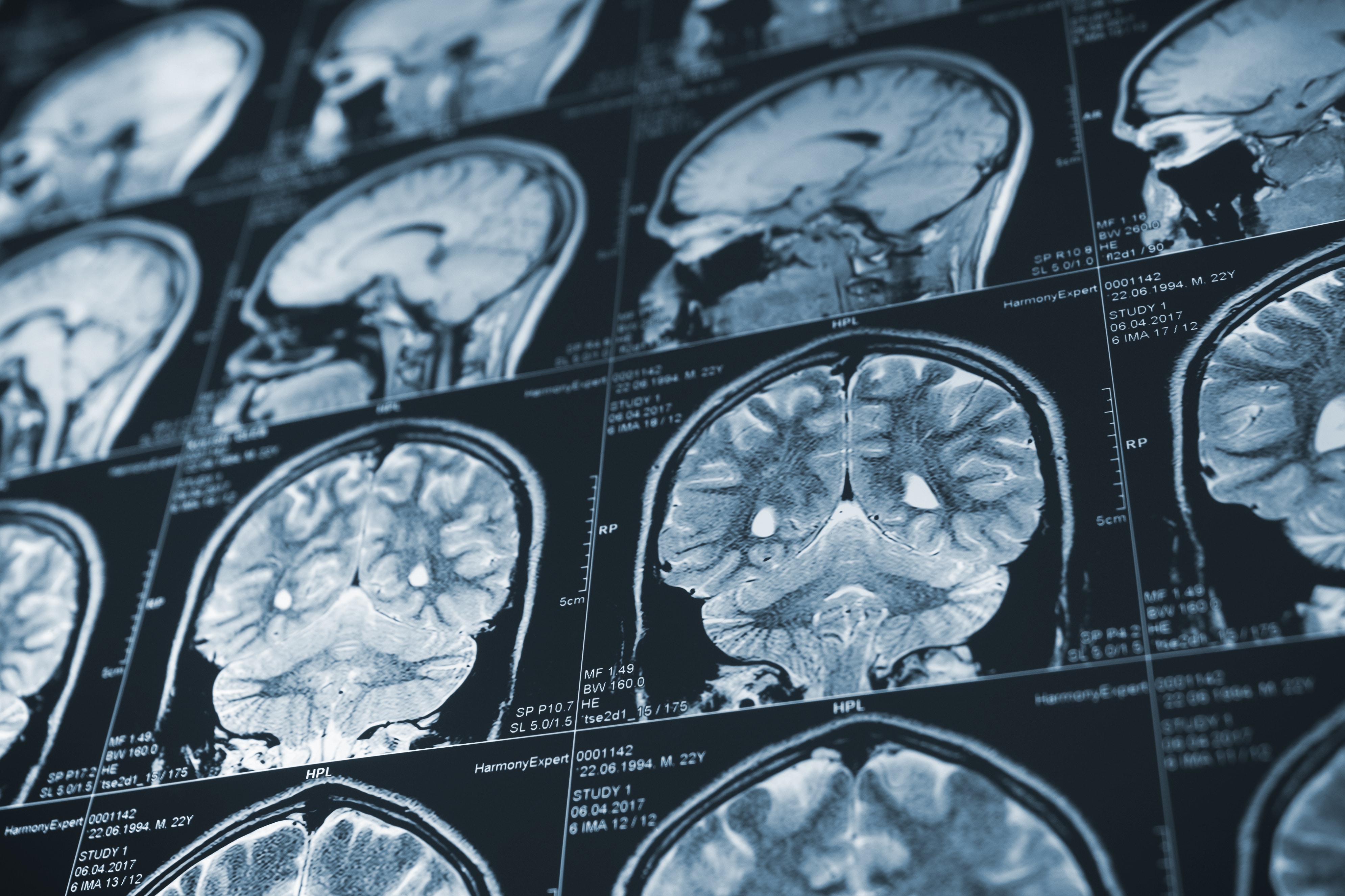
Recently, some new diagnostic tools have been developed which can produce objective signs of permanent brain injury years after the initial trauma. The latest neuro-anatomic technology is a test called the “3 Tesla MRI”, which provides the ability to compare the brain of someone who has had a brain injury with a group of normal control subjects of the same age. Despite the fact that these tests may take place a number of years following the injury, they constitute the most current technology to identify patterns of change and reveal objective evidence of injuries not seen in routine readings of MRI and CT Scan imaging.[3]
The 3 Tesla MRI differs from a standard MRI test in the strength and measurement of the magnetic field. The 3 Tesla MRI allows images to a resolution of a half-millimeter in terms of brain structure. In addition, this new MRI allows for a new very specific type of measurement of the brain tissue to get an objective percentage-based finding of how the brain-injured person’s brain compares to normal brain tissue.
Gregory O’Shanick, MD, an expert in this area, has described it thus, “The technology uses a sophisticated mathematical formula to calculate the volume of the brain based upon measurements of brain tissue with a specific type of imaging sequence.”
The studies are performed using a software program that provides healthcare providers with the means to quantify volume within regions of the brain. The comparisons of measurements are done in percentiles so that the patient would be evaluated in comparison to normal subjects of the same age range. The results would reveal how the injured person’s brain compares, in terms of a percentage of volume, to the normal range. Having this objective evidence using real numbers and percentages would appear to be an invaluable tool in attempting to prove the permanence of a brain injury even a number of years after the injury occurred.
These findings are especially useful when considered in the context of “Iowa Collateral Head Injury Interviews.” This interview format for suspected head injury victims has been proven through numerous studies to be effective in evaluating the nature and extent of the symptoms exhibited by the brain-injured person. Persons interviewed would include close family members and friends. The interviews focus on the behavioral changes exhibited by the brain-injured person. The Iowa Collateral Head Injury Interview test evaluates 21 different items that are ranked from 0 to 2 (0 being no change and 2 being substantial change). Collateral Head Injury Interview test scores at ten or higher are seen as being clearly abnormal.
When the findings of these interviews (the subjective evidence of neuro-behavioral problems) and the objective evidence of the 3 Tesla MRI tests are presented together, they provide a very persuasive means of determining and proving the nature and extent of the brain injury. In the context of civil litigation, this is extremely helpful.[4]
For example, a 60-year-old woman suffering from moderate traumatic brain injury could have a regular MRI, interpreted in the traditional way, which would reveal no change. Using the new technology, the report could reveal any change she experienced as a consequence of the injury which was not detected in the traditional MRI reading.
From a purely neurological and healthcare perspective, these tests are of great importance, and can assist in ensuring the nature and extent of a person’s brain injury is fully recognized and diagnosed, so that treatment can be provided accordingly.
